
The ureter is indicated by its crucial role in the urinary system, serving as a vital conduit for urine transport from the kidneys to the bladder. Understanding its structure, function, and clinical significance is essential for maintaining urinary health.
This article delves into the fascinating world of the ureter, exploring its intricate anatomy, physiological functions, diagnostic techniques, and therapeutic interventions.
Definition and Overview

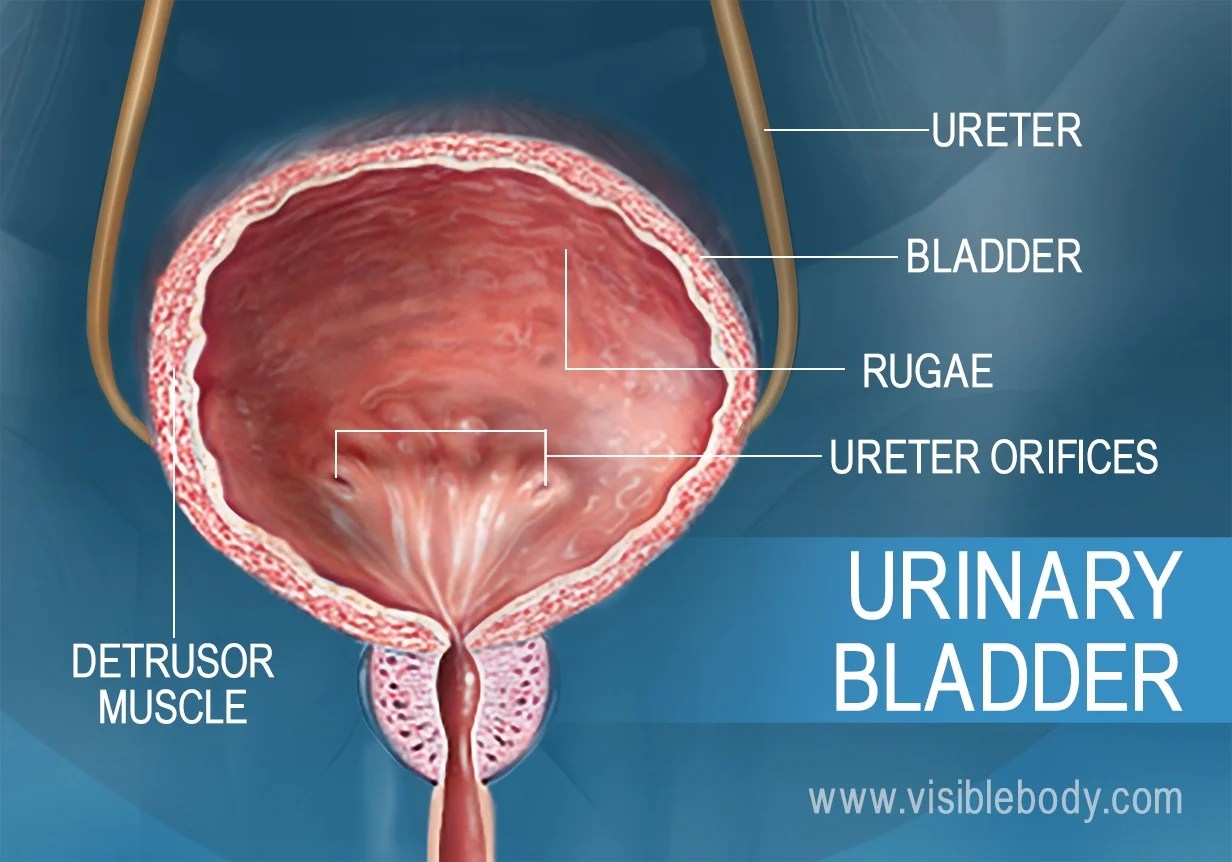
The ureter is a narrow, muscular tube that transports urine from the kidneys to the bladder. It originates from the renal pelvis, the funnel-shaped structure that collects urine from the kidney’s nephrons, and extends to the urinary bladder, where it empties urine through a small opening called the ureteric orifice.
The ureter plays a crucial role in the urinary system by ensuring the efficient flow of urine from the kidneys to the bladder. This process is essential for maintaining fluid balance, removing waste products from the body, and preventing the accumulation of urine in the kidneys.
Anatomical Location
The ureters are located retroperitoneally, meaning they lie behind the peritoneum, the membrane that lines the abdominal cavity. They descend obliquely from the kidneys, passing through the abdomen and pelvis, and entering the urinary bladder at the posterolateral angles of its base.
The ureter is indicated by a tube that transports urine from the kidneys to the bladder. Want to read more about SWOT analysis? Check out Swot Analysis For Toms Shoes to learn more about the topic.
The ureters are approximately 25-30 centimeters in length and have a diameter of about 3-4 millimeters. They are surrounded by a layer of smooth muscle that allows them to contract and propel urine downward.
Structure and Anatomy

The ureter, a crucial component of the urinary system, serves as a conduit for urine from the kidneys to the urinary bladder. Understanding its structure and anatomy is essential for comprehending its function and potential pathological conditions.
The ureter is a muscular tube, approximately 25-30 centimeters in length, originating at the renal pelvis in the kidney and terminating at the urinary bladder. It is composed of three distinct layers: the mucosa, muscularis, and adventitia.
Mucosa
The innermost layer, the mucosa, comprises transitional epithelium, a specialized type of stratified epithelium that lines the urinary tract. It is highly adaptable, allowing for expansion and contraction during urine flow. The mucosa also contains glands that secrete mucus, which aids in lubrication and protection against urinary tract infections.
Muscularis
The muscularis, the middle layer, consists of three layers of smooth muscle: an inner longitudinal layer, a middle circular layer, and an outer longitudinal layer. These muscle layers enable peristaltic contractions, coordinated waves of muscle contractions, which propel urine towards the bladder.
Adventitia, The ureter is indicated by
The outermost layer, the adventitia, is composed of connective tissue and provides structural support to the ureter. It contains blood vessels and nerves that supply the ureter with nutrients and innervation, respectively.
Nerve and Blood Supply
The ureter receives nerve supply from the renal plexus, a network of nerves originating from the lumbar sympathetic chain. These nerves regulate the smooth muscle contractions of the ureter, ensuring efficient urine transport.
The blood supply to the ureter is derived from multiple sources, including the renal artery, abdominal aorta, and common iliac artery. These arteries provide oxygen and nutrients to the ureter, supporting its proper function.
Clinical Significance
The ureter plays a vital role in the urinary system by transporting urine from the kidneys to the urinary bladder. Any abnormalities or disorders affecting the ureter can significantly impact urinary tract function.
Various diseases and conditions can affect the ureter, including:
- Ureteral stones:These are hard mineral deposits that form in the ureter, causing pain, obstruction, and infection.
- Ureteral stricture:A narrowing of the ureter that can obstruct urine flow and lead to hydronephrosis (swelling of the kidney).
- Ureteral tumors:Benign or malignant growths that can develop in the ureter, causing obstruction, bleeding, and pain.
- Ureteropelvic junction obstruction (UPJO):A blockage at the junction where the ureter connects to the renal pelvis, leading to urine buildup and kidney damage.
- Vesicoureteral reflux (VUR):A condition where urine flows backward from the bladder into the ureter, potentially causing kidney infections.
Diagnosing ureteral conditions involves a combination of physical examination, urine analysis, imaging tests (such as X-rays, ultrasound, and CT scans), and cystoscopy (visual examination of the urinary tract using a thin tube with a camera).
Treatment options for ureteral disorders vary depending on the underlying cause and severity. They may include:
- Medical management:Medications to dissolve stones or relieve pain.
- Endoscopic procedures:Using a thin tube with a camera and surgical instruments to remove stones, widen strictures, or repair defects.
- Open surgery:In cases where endoscopic procedures are not feasible or effective.
Imaging and Visualization

Imaging techniques play a crucial role in visualizing the ureters, allowing healthcare professionals to assess their structure, identify abnormalities, and guide treatment decisions. Various imaging modalities offer different perspectives and advantages in ureteral evaluation.
Imaging Modalities
*
-*Ultrasound
Ultrasound uses high-frequency sound waves to create images of the ureters. It is non-invasive, widely available, and relatively inexpensive. Ultrasound can detect dilation, stones, and masses within the ureters.*
-*Intravenous Pyelography (IVP)
IVP involves injecting a contrast agent into a vein, which is then excreted by the kidneys and visualized as it travels through the ureters. IVP provides detailed images of the entire urinary tract, including the ureters, and can reveal blockages, narrowing, or other abnormalities.*
-*Computed Tomography (CT)
CT uses X-rays and computer processing to create cross-sectional images of the body. CT scans can provide high-resolution images of the ureters, allowing for the evaluation of their size, shape, and surrounding structures. CT is particularly useful for detecting ureteral stones and masses.*
-*Magnetic Resonance Imaging (MRI)
MRI uses magnetic fields and radio waves to create detailed images of the body. MRI can provide excellent visualization of the ureters, including their relationship to adjacent organs and structures. MRI is often used to evaluate ureteral injuries, tumors, or congenital anomalies.*
-*Retrograde Pyelography (RPG)
RPG involves inserting a catheter into the ureter through the urethra and injecting a contrast agent. RPG provides direct visualization of the ureteral lumen and can help identify strictures, stones, or other obstructions.The choice of imaging modality for ureteral evaluation depends on the specific clinical situation, the suspected abnormality, and the availability of resources.
These imaging techniques provide valuable information for diagnosing and managing ureteral conditions.
Surgical Interventions

Ureteral surgery is performed to treat a variety of conditions affecting the ureter, including blockages, injuries, and tumors. The type of surgery performed will depend on the specific condition being treated.
Some of the most common types of ureteral surgeries include:
- Ureteroscopy:A minimally invasive procedure that involves inserting a small camera into the ureter to visualize and treat blockages or other abnormalities.
- Ureterolithotomy:A surgical procedure to remove a kidney stone that has become lodged in the ureter.
- Ureteroplasty:A surgical procedure to repair a damaged or narrowed ureter.
- Ureterectomy:A surgical procedure to remove a portion of the ureter.
The outcomes of ureteral surgery are generally good, with most patients experiencing significant improvement in their symptoms. However, as with any surgery, there are some risks involved, including bleeding, infection, and damage to surrounding organs.
Related Structures and Associations: The Ureter Is Indicated By

The ureters are intimately related to various surrounding structures within the abdomen and pelvis, forming close anatomical associations that have clinical implications for both ureteral function and surgical interventions.
Understanding these relationships is crucial for accurate diagnosis, appropriate treatment planning, and minimizing complications during surgical procedures.
Peritoneum
The ureters are retroperitoneal structures, meaning they lie outside the peritoneal cavity. However, they traverse the peritoneal cavity at two points:
- Abdominal cavity:The ureters pass through the posterior abdominal wall at the level of the L4-L5 vertebrae.
- Pelvic cavity:The ureters enter the pelvic cavity through the pelvic brim.
These peritoneal relationships are important because they can influence the spread of infection or malignancy from the abdominal or pelvic cavities to the ureters.
Blood Vessels
The ureters are closely associated with several major blood vessels:
- Abdominal aorta:The right ureter crosses the abdominal aorta anteriorly.
- Inferior vena cava:The left ureter crosses the inferior vena cava posteriorly.
- Gonadal vessels:The ureters are crossed by the gonadal arteries and veins, which supply the testes or ovaries.
These vascular relationships are significant during surgical procedures, as careful dissection is necessary to avoid injury to these vessels.
Lymphatic Vessels
The ureters are drained by lymphatic vessels that follow the course of the blood vessels:
- Abdominal lymph nodes:The lymphatic drainage of the upper ureters goes to the abdominal lymph nodes.
- Pelvic lymph nodes:The lymphatic drainage of the lower ureters goes to the pelvic lymph nodes.
The lymphatic drainage of the ureters is important for understanding the spread of cancer cells and the development of metastases.
Nerves
The ureters are innervated by nerves from the:
- Renal plexus:The upper ureters are innervated by the renal plexus.
- Pelvic plexus:The lower ureters are innervated by the pelvic plexus.
These nerve connections provide sensory innervation to the ureters and allow for involuntary control of ureteral peristalsis.
Developmental Aspects
The ureter, a crucial component of the urinary system, originates from a complex embryological journey. During the fourth week of gestation, a structure called the Wolffian duct, initially responsible for forming the male reproductive system, plays a pivotal role in ureter development.
The upper portion of the Wolffian duct gives rise to the ureteric bud, which descends caudally, meeting the metanephric blastema, the precursor to the kidney. This interaction triggers the formation of the ureter and the renal collecting system.Congenital anomalies of the ureter are not uncommon, resulting from disruptions during this intricate developmental process.
These anomalies can manifest in various forms, including ureteral duplication, where two ureters drain a single kidney; ectopic ureter, where the ureter inserts into an abnormal location; and ureterocele, a cystic dilation of the distal ureter within the bladder.Understanding the embryological basis of ureteral development is essential for comprehending the clinical implications of these congenital disorders.
Accurate diagnosis and appropriate management, often involving surgical interventions, are crucial for preserving urinary function and preventing complications.
Clinical Implications and Management of Ureteral Developmental Disorders
Congenital anomalies of the ureter can lead to a spectrum of clinical presentations, ranging from asymptomatic findings to severe urinary tract infections, hydronephrosis, and even renal failure. The management of these disorders depends on the specific anomaly, its severity, and the presence of associated complications.In
cases of ureteral duplication, surgical intervention may be necessary to correct the abnormal drainage pattern and prevent urinary stasis. Ectopic ureters often require reimplantation into the bladder to establish proper urinary flow. Ureterocele, if causing significant obstruction or recurrent infections, may be treated with endoscopic incision or open surgical excision.Regular
monitoring and follow-up are essential for individuals with congenital ureteral anomalies to detect and manage any potential complications promptly. Early diagnosis and appropriate treatment can significantly improve outcomes and preserve renal function in these patients.
Key Questions Answered
What is the primary function of the ureter?
The ureter’s primary function is to transport urine from the kidneys to the bladder, facilitating its elimination from the body.
What are the common diseases affecting the ureter?
Ureteral diseases include urinary tract infections, kidney stones, and ureteral strictures, which can obstruct urine flow and cause discomfort.
How is the ureter visualized for diagnostic purposes?
Imaging techniques such as ultrasound, X-rays, and CT scans are commonly used to visualize the ureter and assess its structure and function.
